This is not merely a clinical description; it is the unfolding story of an infection, from its microscopic origins to its profound impact on the human body and psyche. It is a tale of a bacterial invader with a particular affinity for the lymphatic system, a narrative marked by stages of stealth, inflammation, and, if left unchecked, devastating chronic complications. For a knowledgeable audience, understanding LGV demands a journey through its historical context, its unique microbiological characteristics, the distinct chapters of its clinical manifestation, its epidemiological resurgence, and the modern approaches to diagnosis, treatment, and prevention.
The Unseen Architect: Chlamydia trachomatis Serovars L1, L2, L3
At the heart of LGV lies a bacterium, Chlamydia trachomatis. However, this is not the common Chlamydia that causes the vast majority of genital and ocular infections worldwide. Chlamydia trachomatis is a species with multiple distinct serovars, each possessing a unique tropism and pathogenic profile. While serovars D through K are responsible for ocular trachoma and the more common urogenital chlamydial infections, it is the specific serovars L1, L2, and L3 that orchestrate the development of LGV.
These "L" serovars are distinguished by their enhanced invasiveness. Unlike their counterparts, which primarily colonize mucosal surfaces, L1, L2, and L3 possess a greater capacity to penetrate epithelial cells, replicate, and disseminate beyond the superficial layers. Their particular predilection for lymphatic tissue is the defining feature that sets LGV apart. This invasive capability allows the bacteria to travel swiftly from the initial site of infection to regional lymph nodes, initiating a cascade of inflammation and destruction that characterizes the disease’s progression.
To truly appreciate the bacterial architect, one must briefly acknowledge the unique life cycle of Chlamydia. It is an obligate intracellular bacterium, meaning it cannot reproduce outside a host cell. Its existence oscillates between two distinct forms: the elementary body (EB) and the reticulate body (RB). The EB is the infectious, metabolically inert form, designed for extracellular survival and host cell entry. Once inside a cell, the EB transforms into the RB, a metabolically active, non-infectious form that replicates profusely. After numerous divisions, RBs reorganize back into EBs, which are then released to infect new cells, perpetuating the cycle of invasion and replication. In LGV, this cycle occurs within macrophages and other immune cells, facilitating its spread through the lymphatic system.
A Tapestry of Time: LGV’s Historical Footprint
The story of LGV is not a new one. It has been recognized, though not always fully understood, for centuries. Early descriptions of what was likely LGV can be traced back to the time of Hippocrates, with accounts of inguinal swellings and genital ulcers. However, it was only in the early 20th century that a clearer picture began to emerge. In 1913, Durand, Nicolas, and Favre provided a seminal description of a "subacute lymphogranulomatosis," which laid the groundwork for modern understanding. They characterized the distinct clinical presentation, particularly the inguinal lymphadenopathy that became a hallmark of the disease.
For much of the 20th century, LGV was considered one of the "classic" venereal diseases, often referred to as the "fourth venereal disease" after syphilis, gonorrhea, and chancroid. It was prevalent in tropical and subtropical regions, particularly in parts of Africa, Asia, and South America, where it was endemic. In these regions, the devastating chronic complications, such as genital elephantiasis and rectal strictures, were not uncommon. However, with the advent of effective antibiotics in the mid-20th century, particularly tetracyclines, and improvements in public health infrastructure, the incidence of LGV began to decline significantly in many parts of the world, leading to its gradual relegation to a "rare" or "forgotten" disease in developed nations. Clinicians trained in recent decades might only have encountered LGV in textbooks, a historical footnote rather than a pressing clinical concern.
The Narrative of Manifestation: Clinical Stages
The story of LGV within an infected individual unfolds in distinct, though sometimes overlapping, stages, each revealing a deeper penetration and broader impact of the bacterial invader.
1. The Primary Lesion: A Fleeting Whisper (Incubation: 3-30 days)
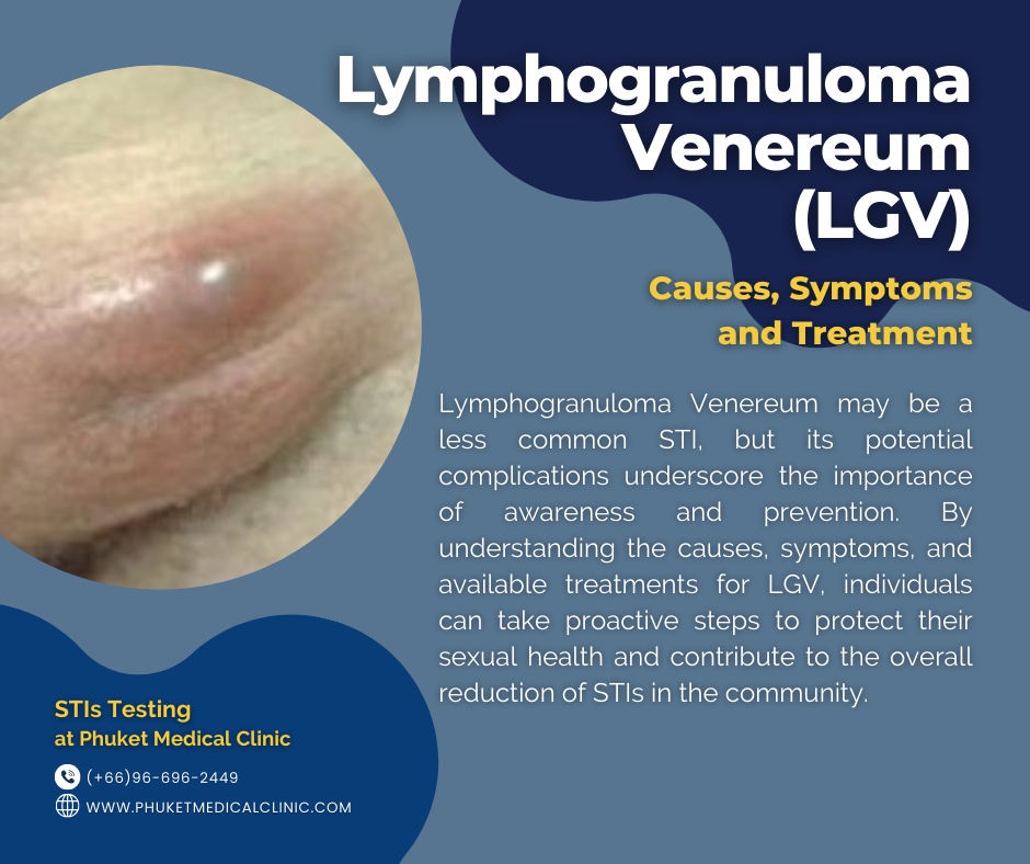
The initial chapter of LGV often begins with a subtle, easily overlooked event: the primary lesion. After the bacteria penetrate the skin or mucous membranes at the site of inoculation (genital, anal, or oral), a small, painless papule, vesicle, or ulcer (chancre) may develop. This lesion is typically transient, often healing spontaneously within a few days and frequently going unnoticed by the infected individual. Its innocuous nature is deceptive, as it marks the entry point for the more profound lymphatic invasion that follows. Because it is painless and resolves quickly, patients rarely seek medical attention at this stage, contributing to delayed diagnosis. In some cases, particularly with oral or anal inoculation, the primary lesion may be entirely internal and asymptomatic.
2. The Lymphatic Drama: Swelling and Systemic Stirrings (2-6 weeks after exposure)
This is the most characteristic and often dramatic chapter of LGV. The bacteria, having breached the initial barrier, journey to the regional lymph nodes, where they multiply and incite a vigorous inflammatory response.
-
Inguinal Lymphadenopathy (Buboes): The classic presentation involves the enlargement and inflammation of the inguinal lymph nodes, typically unilateral but sometimes bilateral. These swollen nodes, known as buboes, become firm, tender, and eventually fluctuant as they suppurate and form abscesses. The overlying skin may become inflamed, red, and edematous. In severe cases, buboes can rupture, discharging pus and necrotic tissue, leading to chronic draining sinuses and fistulas. A historically described, though less common, sign is the "groove sign," where enlarged inguinal lymph nodes are separated by a depression formed by the inguinal ligament, creating a characteristic "groove." This phenomenon highlights the extent of lymphatic involvement both above and below the ligament.
-
Pelvic and Perirectal Lymphadenopathy: In cases of anogenital inoculation, particularly through receptive anal intercourse, the lymphatic drainage involves the perirectal and deep pelvic lymph nodes. This can lead to significant proctitis and proctocolitis, often without prominent external inguinal buboes. The deeper location of these affected nodes makes diagnosis more challenging and the resulting symptoms more severe.
-
Systemic Symptoms: As the lymphatic system becomes overwhelmed, the infection can trigger systemic responses. Patients may experience fever, chills, malaise, headache, anorexia, myalgia, and arthralgia. These constitutional symptoms reflect the body’s generalized inflammatory reaction to the disseminating infection. Occasionally, hepatosplenomegaly or meningitis can occur, indicating a more widespread systemic involvement.
3. The Chronic Aftermath: A Scarred Landscape (Months to Years)
If LGV remains untreated or is inadequately managed, the story progresses to a devastating chronic phase, leaving behind a scarred and often disfigured landscape. This tertiary stage is particularly debilitating and contributes significantly to long-term morbidity.