In the vast, intricate tapestry of human health, some threads are vibrant and obvious, demanding immediate attention. Others, however, are woven subtly, almost invisibly, into the fabric, their presence often underestimated or entirely overlooked until their cumulative strain begins to unravel the whole. Among these silent, pervasive forces is trichomoniasis, an infection caused by the microscopic parasite Trichomonas vaginalis. While often dismissed as a benign nuisance, particularly when asymptomatic, the story of trichomoniasis is far more complex and compelling than its understated reputation suggests. It is a narrative of a ubiquitous pathogen, a hidden epidemic, and a profound, yet frequently unacknowledged, impact on global public health, individual well-being, and even the very fabric of human relationships.
This article embarks on a journey to tell that story, delving deep into what trichomoniasis is, how it operates, and the multifaceted ways it affects our health – often in concert with other, more loudly proclaimed health challenges. For a knowledgeable audience, we will move beyond superficial descriptions to explore the biological intricacies of the parasite, the spectrum of its clinical manifestations, the far-reaching complications it precipitates, the evolving landscape of its diagnosis and treatment, and the critical public health implications that demand our urgent attention.
Chapter 1: The Architect of Disease – Trichomonas vaginalis Revealed
To understand trichomoniasis, we must first understand its protagonist: Trichomonas vaginalis. This single-celled, flagellated protozoan is a fascinating, albeit insidious, organism. Unlike many other sexually transmitted infections (STIs) caused by bacteria or viruses, T. vaginalis is a parasite, belonging to the phylum Metamonada, specifically the order Trichomonadida. It is remarkably adapted to its human host, having forgone a free-living stage or a cyst form, existing solely as a trophozoite – the active, infective stage.
1.1 Morphology and Biology: A Master of Adaptation
The trophozoite of T. vaginalis is typically pear-shaped (pyriform), ranging from 7 to 30 micrometers in length, making it slightly larger than a white blood cell. Its defining features include:
- Flagella: Four anterior flagella, responsible for its characteristic jerky, tumbling motility, and one posterior flagellum that forms the outer border of an undulating membrane, aiding in adhesion and movement.
- Axostyle: A prominent, rod-like structure extending through the length of the organism, providing structural support and potentially involved in host cell attachment.
- Hydrogenosome: A unique, membrane-bound organelle that functions similarly to mitochondria but in anaerobic conditions. This adaptation allows T. vaginalis to thrive in the low-oxygen environment of the vaginal canal and male urethra, fermenting pyruvate to acetate, CO2, and hydrogen. This metabolic pathway is crucial for its survival and offers a key target for certain therapeutic drugs.
- Adhesion Factors: The parasite possesses an array of surface molecules, including adhesins, lipophosphoglycan (LPG), and cysteine proteinases, which facilitate its attachment to epithelial cells of the urogenital tract. These adhesion mechanisms are vital for colonization and evading host immune responses.
1.2 Lifecycle: A Direct and Efficient Pathogen
The lifecycle of T. vaginalis is remarkably simple and direct, contributing to its efficient transmission. There is no intermediate host or environmental stage. Transmission occurs almost exclusively through direct contact, primarily sexual intercourse. Once transmitted, the trophozoites multiply by binary fission in the urogenital tract – the vagina, urethra, and prostate being the primary sites of infection. They feed on bacteria, host cells, and cellular debris. The absence of a cyst stage, which would typically provide environmental resilience, highlights its complete dependence on direct host-to-host transfer and explains its relative fragility outside the human body. However, it can survive briefly on moist surfaces, such as towels or toilet seats, though this mode of transmission is considered rare.
Understanding these biological intricacies provides the foundation for comprehending the parasite’s persistence, its resistance to certain treatments, and the challenges inherent in its diagnosis and eradication.
Chapter 2: The Clinical Manifestations – A Spectrum of Silence and Suffering
One of the most defining and problematic characteristics of trichomoniasis is its often asymptomatic nature. This "silent" aspect is central to its widespread prevalence and contributes significantly to its underestimation and continued transmission. However, when symptoms do emerge, they can range from mild irritation to severe, debilitating discomfort. The clinical presentation often differs between sexes, further complicating the diagnostic picture.
2.1 Symptoms in Women: A Common Urogenital Complaint
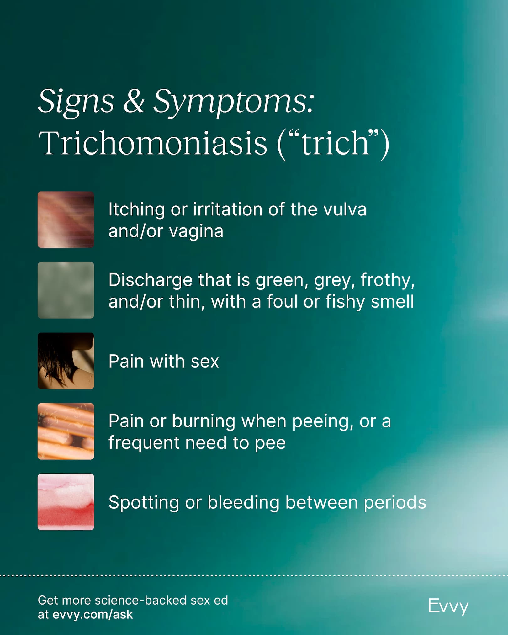
Women are more likely than men to experience symptomatic infection, though estimates suggest that up to 70-85% of infected women remain asymptomatic. When symptoms occur, they typically manifest as:
- Vaginitis: Inflammation of the vagina, often leading to a characteristic discharge.
- Vaginal Discharge: This is the hallmark symptom, frequently described as frothy, greenish-yellow, and malodorous (often with a "fishy" smell, particularly after intercourse due to the interaction with seminal fluid). The frothiness is attributed to the gas produced by the parasite’s metabolic activity.
- Vulvar Itching and Irritation: Intense pruritus (itching) and burning sensation in the vulva and vagina.
- Dysuria: Pain or burning during urination, often mistaken for a urinary tract infection (UTI).
- Dyspareunia: Pain during sexual intercourse.
- Abdominal Discomfort: Lower abdominal pain can occur in more severe cases.
- "Strawberry Cervix" (Colpitis Macularis): A classic, though uncommon (seen in 2-5% of cases), sign observed during speculum examination. This appearance is due to punctate hemorrhages on the cervical mucosa, giving it a mottled, erythematous (reddened) appearance.