In the vast and intricate tapestry of human ailments, some diseases exist in the bright glare of public awareness, their names uttered with familiarity and their symptoms widely recognized. Others, however, lurk in the shadows, emerging periodically to remind us of their persistent, often devastating, presence. Lymphogranuloma Venereum (LGV) is one such entity – a silent infiltrator, a master of disguise, and a forgotten foe that has recently re-emerged from the annals of medical history to challenge our diagnostic acumen and public health strategies.
To speak of LGV is to embark on a journey through microbiology, epidemiology, clinical medicine, and social history. It is a story of a cunning pathogen, Chlamydia trachomatis, and its particularly virulent serovars, which orchestrate a multi-stage drama within the human body, often culminating in profound suffering and long-term complications. For a knowledgeable audience, understanding LGV requires delving beyond surface symptoms, into the very cellular mechanisms of infection, the nuances of its clinical presentation, and the complex interplay of societal factors that have fueled its recent resurgence.
The Protagonist: Chlamydia trachomatis and its L-Serovars
Our story begins with the pathogen itself: Chlamydia trachomatis. This seemingly innocuous bacterium is an obligate intracellular parasite, meaning it cannot replicate outside of a host cell. It has a unique biphasic developmental cycle, alternating between two distinct forms:
- Elementary Body (EB): The infectious, metabolically inert form. These small, resilient particles are specialized for extracellular survival and attachment to host cells.
- Reticulate Body (RB): The non-infectious, metabolically active, and replicative form. Once inside the host cell, EBs differentiate into RBs, which multiply rapidly within a membrane-bound vacuole called an inclusion.
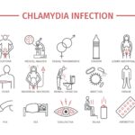
Most C. trachomatis infections are well-known, causing common sexually transmitted infections (STIs) like urethritis, cervicitis, and conjunctivitis, as well as trachoma (a leading cause of preventable blindness). These are typically caused by serovars A-K. However, the villains of our LGV narrative are a distinct subgroup: serovars L1, L2, and L3.
What sets the L-serovars apart, making them capable of such systemic devastation, is their enhanced invasive capacity. Unlike their A-K cousins, which tend to remain localized to mucosal surfaces, L-serovars possess a greater tropism for lymphatic tissue and can traverse mucosal barriers more effectively. This allows them to spread beyond the initial site of infection, gaining access to the lymphatic system, where they wreak havoc, orchestrating the characteristic pathological changes that define LGV. This ability to invade and disseminate is the crucial plot twist that elevates LGV from a simple mucosal infection to a systemic disease with potentially crippling consequences.
The Unfolding Drama: Stages of LGV
The clinical presentation of LGV is a multi-act play, unfolding over weeks, months, or even years, and often characterized by a deceptive initial silence followed by dramatic, painful manifestations.
Act I: The Primary Lesion – A Fleeting Whisper
The incubation period for LGV typically ranges from 3 to 30 days, averaging around 7-12 days. The initial event, the primary lesion, is often the most overlooked and understated act of the entire drama. It manifests at the site of inoculation as a small, painless papule, vesicle, or ulcer (chancre). Crucially, this lesion is typically:
- Transient: It often heals spontaneously within a few days, sometimes even before the individual is aware of its presence.
- Painless: Its lack of discomfort contributes significantly to its being missed or dismissed.
- Small and Atypical: It may be easily confused with other minor skin irritations or go completely unnoticed, especially if located internally (e.g., within the rectum or urethra).
The location of this primary lesion is dictated by the mode of transmission. In heterosexual individuals, it is commonly found on the penis, scrotum, labia, vaginal wall, or cervix. In men who have sex with men (MSM), anorectal inoculation is increasingly common, leading to primary lesions within the rectum or anal canal, which are almost invariably asymptomatic and undetectable.
The fleeting nature of this primary lesion is a significant diagnostic challenge. Patients rarely present during this stage, meaning healthcare providers are almost always confronted with the disease in its more advanced, secondary manifestations. This initial "whisper" often goes unheard, setting the stage for the more explosive secondary act.
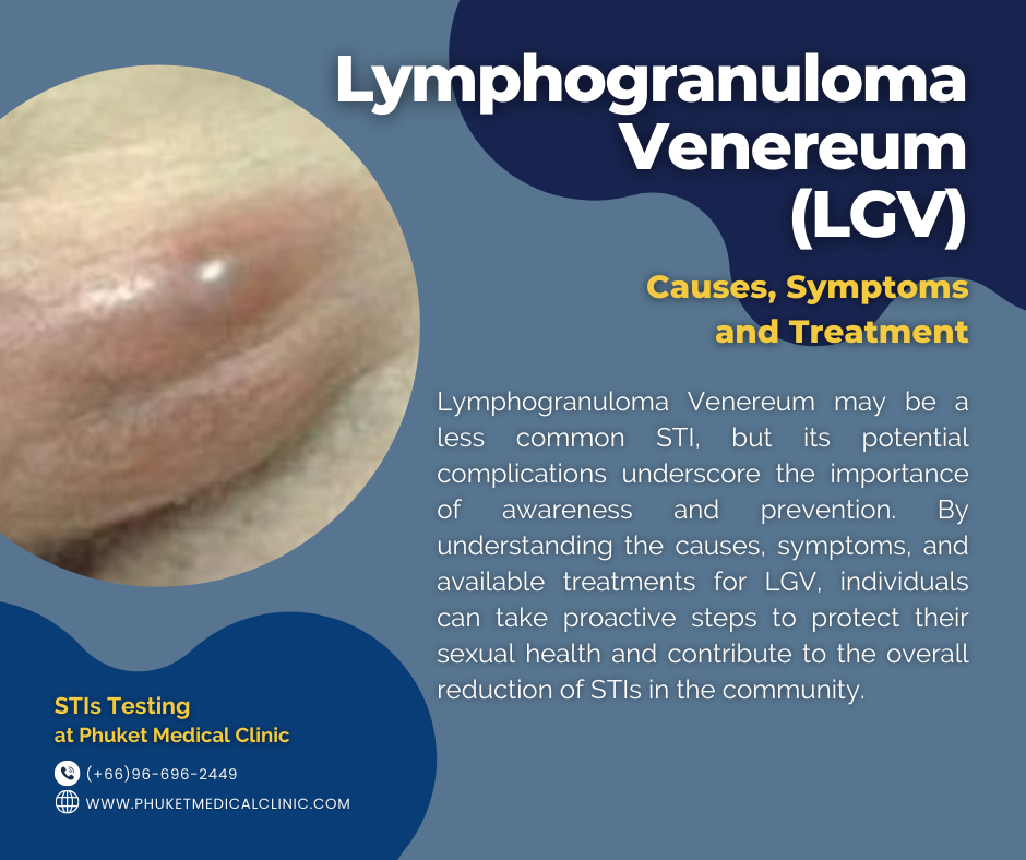
Act II: The Secondary Stage – The Lymphatic Battleground
Weeks after the primary lesion has healed (typically 1 to 4 weeks, but sometimes longer), the disease enters its secondary, or "inguinal syndrome," stage. This is where the L-serovars, having silently invaded the lymphatic system, begin their overt assault. The lymphatic vessels draining the initial site of infection become inflamed, and the regional lymph nodes swell and become exquisitely tender.
The clinical presentation of this secondary stage varies depending on the site of primary inoculation:
-
Inguinal Syndrome (Genital/Groin LGV): This is the classic presentation, historically associated with LGV.
- Lymphadenopathy: Unilateral or bilateral inguinal lymph node swelling is characteristic. These nodes can become remarkably enlarged, firm, and painful.